Human Skeleton System: Structure and Functions
Every object in the world depends on its structure for strength and stability. A building stands because of its pillars and beams, a vehicle runs because of its chassis, and the human body survives and moves because of its skeleton. The human skeleton may sound scary to some people because of how it is shown in movies, but in reality, it is simply the internal framework of our body. Without the skeleton, the human body would not have shape, strength, or the ability to move. Understanding the human skeleton helps us understand how our body works and how it protects our vital organs.
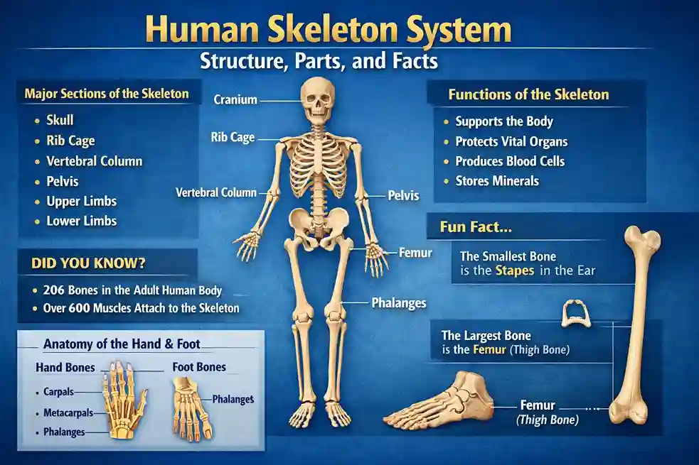
The human skeleton is also called the musculoskeletal system. It forms the internal structure of the body and protects important organs such as the brain, heart, lungs, liver, and kidneys. It also supports movement by working together with muscles, joints, and ligaments. An adult human body has 206 bones, while children are born with around 300 bones. As children grow, some bones fuse together to form stronger structures. Most of the calcium present in the human body is stored in bones, which helps maintain their strength and density.
Basic Features of the Human Skeleton
The longest bone in the human body is the femur, also known as the thigh bone. It is very strong and supports most of the body’s weight. The smallest bone is the stirrup bone, which is located inside the ear. Bones are not lifeless structures; they are living tissues that grow, repair themselves, and change throughout life.
The human skeleton is divided into two main parts: the axial skeleton and the appendicular skeleton. Some classifications also include a smaller group related to special bones, but the axial and appendicular skeletons are the main divisions.
Axial Skeleton and Its Components
The axial skeleton forms the central axis of the body. It includes the skull and the vertebral column. These structures protect the brain and spinal cord, which are the most important parts of the nervous system.
The skull is the most important part of the axial skeleton because it protects the brain. This is why wearing a helmet is necessary while riding a two-wheeler. The upper part of the skull, located above the nose, is called the cranium. The cranium acts as a protective case for the brain. It is made of two types of bone formation: cartilaginous and membranous. The cartilaginous part forms the base of the skull, while the membranous part forms the sides and roof.
The bones around the eyes are called orbital or zygomatic bones. The upper jaw bone is known as the maxilla and remains fixed in position. The lower jaw bone is called the mandible, which is movable and helps in chewing and speaking. In newborn babies, the skull bones are not fully fused. This flexible structure helps the baby pass through the birth canal safely. Over time, these bones fuse and become stronger. The skull includes major bones such as the frontal, temporal, and occipital bones. Breaking the skull requires a very high amount of force, which shows how strong it is.
Vertebral Column and Its Structure
The vertebral column, also known as the spine or backbone, acts like a natural spring in the human body. It has an S-shaped curve, which helps absorb shocks when we walk or run on hard surfaces. This shape allows flexibility and balance.
The vertebral column is divided into five regions. The first region is the cervical region, which includes seven bones labeled C1 to C7. These bones are located in the neck area. The second region is the thoracic region, consisting of twelve bones from T1 to T12. The third region is the lumbar region, which has five bones labeled L1 to L5. The fourth region is the sacral region, made of five fused bones. The last region is the coccygeal region, consisting of four small fused bones.
Each individual bone of the vertebral column is called a vertebra. A vertebra has several parts, including the body, spinous process, transverse process, vertebral arch, and vertebral foramen. The spinal cord passes through the vertebral canal, which protects it from injury. Small joints called facets prevent bones from rubbing against each other.
The cervical vertebrae include two special bones called the atlas and axis. These bones allow the head to move up, down, and side to side. The atlas supports the skull, while the axis helps rotate the neck.
Appendicular Skeleton and Its Functions
The appendicular skeleton connects the limbs to the axial skeleton. It plays an important role in movement, weight transfer, and support. It helps attach the upper and lower limbs to the body and transfers the weight of the upper body to the lower limbs. It also protects organs in the pelvic region and provides attachment points for muscles.
The shoulder region is called the pectoral girdle. It includes the scapula, which is a triangular bone behind the shoulder, and the clavicle, also known as the collarbone. These bones help move the arms and support the upper chest.
The upper arm bone is called the humerus. Below it are two bones in the forearm: the radius and the ulna. The radius is on the thumb side, while the ulna is on the other side. The wrist contains small bones called carpals. The palm has five bones called metacarpals, and the fingers have bones called phalanges.
Pelvic Girdle and Lower Limbs
The pelvic girdle connects the axial skeleton to the lower limbs. It supports body weight and protects organs such as the urinary, digestive, and reproductive systems. The pelvic girdle is made of three main bones: the ilium, ischium, and pubis. The ischium is also called the sitting bone because it bears weight while sitting.
In females, the pelvic region is wider and lighter to help during childbirth. This type of pelvis is called the gynecoid pelvis and has a wider opening and a larger angle.
The lower limbs include the femur, patella, tibia, and fibula. The femur is the strongest and longest bone. The patella is the kneecap, which protects the knee joint. Below the knee, the tibia is the main weight-bearing bone, while the fibula is thinner and provides support.
The ankle region contains seven bones called tarsals. The foot arch is formed by metatarsals, which act as a bridge between tarsals and toes. The toes are made of phalanges, similar to the fingers.
Important Facts About Human Bones
Bones contain a spongy tissue called bone marrow, which is found mainly in large bones like the pelvis and femur. Out of 206 bones in the human body, 106 are present in the hands and feet alone. The knee joint is the largest joint because it connects three bones and supports body weight.
Teeth are even stronger than bones. The outer layer of teeth is called enamel, which protects the inner nerves and tissues. Bones have a natural healing ability. When a fracture occurs, the body produces new bone cells to repair the damage.
Calcium is essential for healthy bones. Foods like milk, cheese, yogurt, tofu, seeds, beans, leafy vegetables, and nuts help maintain bone strength. Regular exercise, especially weight-bearing activities, also keeps bones strong. A condition called osteoporosis weakens bones with age, making them fragile and prone to fractures.
Conclusion
The human skeleton is the foundation of the body. It provides shape, protection, movement, and strength. From the skull to the toes, every bone has a specific role. Taking care of bones through proper nutrition, exercise, and awareness is essential for a healthy life. The skeleton truly acts as the building blocks of the human body and supports us throughout our lives.